To the inner surface of the heart. The Adams apple is located on.

The part of the serous membrane attached to the fibrous membrane is called the parietal pericardium while the part of the serous membrane attached to the heart is known as the visceral pericardium.
Membrane on the surface of the heart. The serous membrane or serosal membrane is a thin membrane that lines the internal body cavities and organs such as the heart lungs and abdominal cavity. The serous membrane allows for frictionless movement in a number of vital organs. The pericardium of the human heart is a membranous sac that surrounds and protects the heart.
Find how it is divided its function and disorders. The serous membrane that is on the surface of the heart muscle is called the visceral pericardium or epicardium The function of the serous fluid produced by the serous layer is. Layers and Membranes of the Heart.
Terms in this set 5 Endocardium. Innermost layer of cells that lines the atria ventricles and heart valves. Muscular layer of the heart.
This membrane surrounds the heart as the pericardial sac and secretes pericardial fluid. The two surface membranes of rat heart muscle mitochondria are structurally very different. They both also differ structurally from the crista membranes.
There is therefore no justification to consider the crista membranes as mere infoldings of the innermost surface membrane. Most of the earlier freeze-fracture studies on intact mitochondria have predominantly involved the innermost. The endocardium is a thin smooth membrane which lines and gives the glistening appearance.
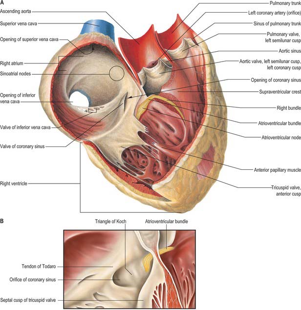
To the inner surface of the heart. It assists in forming the valves by its reduplications and is continuous with the lining membrane of the large bloodvessels. It consists of connective tissue and elastic fibers and is attached to the muscular structure by loose elastic tissue which contains bloodvessels and nerves.
This is lined by a double inner membrane called the serous membrane that produces pericardial fluid to lubricate the surface of the heart. The part of the serous membrane attached to the fibrous membrane is called the parietal pericardium while the part of the serous membrane attached to the heart is known as the visceral pericardium. In actual clinical practice the borders of the heart refer to the borders of its sternocostal Anterior surfaceIt is these same borders that project to the anterior chest wall as the surface projection of the heartThe borders are.
This is slightly convex to the right. It is formed by the right atrium. This is formed by the left ventricle and the left auricle.
Short proteins called cadherins in the plasma membrane connect to intermediate filaments to create desmosomes. The cadherins join two adjacent cells together and maintain the cells in a sheet-like formation in organs and tissues that stretch like the skin heart and muscles. The membrane that directly surrounds the heart and defines the pericardial cavity is called the pericardium or pericardial sac.
It also surrounds the roots of the major vessels or the areas of closest proximity to the heart. A coupled SYSTEM of intracellular Ca2 clocks and surface membrane voltage clocks controls the timekeeping mechanism of the hearts pacemaker. Ion channels on the surface membrane of sinoatrial nodal pacemaker cells SANCs are the proximal cause of an action potential.
Serous membrane attached to lung surface. Serous membrane attached to thoracic wall. Double layered membrane that holds abdominal organs in place.
Double layered membrane that surrounds the heart. The ear is located on. The Adams apple is located on.
What is the membrane on the surface of a lung called. Asked by Wyatt Williams Last updated. May 05 2021 Answer.
Answered Feb 12 2017. I feel like this question should be more clearly asked there is an outer parietal cavity and then an inner visceral. The respiratory chain is located in the inner membrane of mitochondria and produces the major part of the ATP used by a cell.
The heart surface is modelled by means of a thin or thivibrating membrane in order to ck represent the epicardial surface or the connected epicard and myocard. The membrane motion is described by means of a system of coupled linear partial differential equations PDEs whose 3D-input function is assumed to be known. After spatial discretization of the PDE solution space by the Finite Spectral.